Written by Wendy Murdoch
How many times have you cleaned your horse’s feet without thinking about it? You take the hoof pick and remove all the dirt, making sure there are no rocks caught between the hoof, the sole and the shoe. While you were cleaning out that hoof, did you ever consider what your comparable body part is? You might be surprised that you are standing on it. Well almost!

If you were a ballerina, your stance would be more like that of the horse, up on your toes. In fact, the modern horse stands on only one of his toes. Often considered to be the remains of the horse’s prehistoric third toe, the horse’s hoof has evolved to be a unique structure designed to allow your horse swift movement over uneven ground.
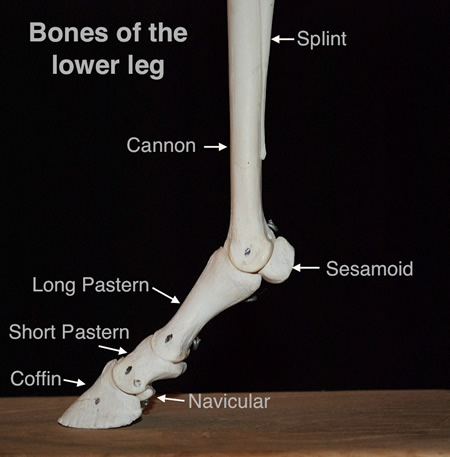
There is some speculation about what happened to the other 4 toes, which the horse’s ancestors may have had. It is generally agreed that two of the toes became the splint bones, which run alongside the cannon bone above the horse’s fetlock. One of the other toes is thought to have become the chestnut, a horny growth inside and above your horse’s knee and inside and below your horse’s hocks. However, some people think that the chestnut may have come from keratinized (fingernail-like) glands that excreted foul-smelling goo or secreta. Secreta is a fluid discharged from a gland. You may have secreted tears when your horse accidentally stepped on your foot even with your boots on!
The first of the other toes of the horse is believed to be the ergot, located on the back of your horse’s fetlock joint. Oftentimes it is surrounded by feathers, long hairs extending from the back of the horse’s lower leg and fetlock. When trimming this area, you have to be careful to go around the ergot. The origin of the word ergot is unknown, but is believed to be derived from a French word, argot, meaning “cock’s spur.” If you have ever seen a rooster, I am sure you will remember the long sharp spurs protruding from the inside of his legs. These very sharp spurs can cause serious damage if you aren’t careful! Since the horse’s ergots protrude from the fetlock, it seems logical to call them ergots.
To recap, the horse stands on your middle toe, two of his toes became splint bones, one became the ergot and one may have become the chestnut. But how about the hoof itself? What are those structures related to in your body?
It is pretty easy to imagine that the hoof wall or capsule is similar to your toenail. In fact, the material your toenail and the hoof wall are made of is the same and is called is keratin, a fibrous protein forming the main structural constituent of hair, feathers, hoofs, claws, horns, etc. The word keratin comes from the Greek work keras, meaning horn.
Underneath the keratin is the laminae, a thin layer, plate or scale of sedimentary rock, organic tissue, or other material. Laminae can be found in many things including your driver’s license, which is laminated between two pieces of plastic to protect it. In the horse’s hoof the laminae is a thin layer of tissue that runs parallel with the hoof wall and is key to the strength and health of the hoof. We have a similar structure under our toenails called the nail bed, which contains blood vessels, nerves and nail-producing cells.
Each of your fingers and toenails has a cuticle, the place where your nail meets your skin. The cuticle fuses the nail to the skin, providing a protective barrier. If the cuticle gets badly damaged, it can affect the growth of your nails. The equivalent of the cuticle in the horse is the coronary or coronet band.
A coronet is a small crown worn by princes and princesses and by other nobles below the rank of sovereign. If you look at your horse’s coronet band you can see why it might be considered a small crown. And given the very old expression “no hoof, no horse” it makes sense to view the coronet band, the source of the hoof wall, as something very important! The coronet band runs along the upper margin of the horse’s hoof, where the hoof and the skin meet. It is important to avoid any damage to the coronet band, as it may damage subsequent hoof growth.
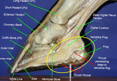
On the bottom and at the back of the hoof are a number of structures: the sole of the foot, the bars, the digital cushion, the bulbs of the heel, and the frog. These specialized structures are designed to provide support, absorb concussion and provide sensory feedback. We don’t have any directly related parts to our fingers and toes; however, we could think of the fleshy part of our finger below the nails as equivalent to these structures.
While the protective function of the fingernail is commonly known, there is a sensory function that is equally important. The fingertip has many nerve endings which allow us to receive volumes of information about objects we touch. The nail acts counter to the fingertip, providing even more sensory input. The horse also receives sensory information through the hoof that helps him adjust his balance. We are still learning about how the horse’s foot handles the concussion of walking and running on this single toe.
Dr. Robert Bowker, a Michigan State University College of Veterinary Medicine researcher, has pieced together a new picture of equine foot physiology that suggests vascular systems in horse hooves function in much the same way that air- or gel-filled running shoes do. “Moving liquids are the best way to dissipate energy. That is why some of the major running shoe manufacturers market products that contain liquids in their soles.”
Bowker’s hypothesis suggests a negative pressure is actually created by the outward movement of the hoof cartilage. This movement creates a vacuum action that sucks blood from beneath the coffin bone into the rear portion of the hoof. “As the blood moves to the rear of the hoof, it dissipates the energy caused by its impact on the ground, much like fluid-filled running shoes do.” So the next time you go running in your gel Nikes imagine your running shoes are the shock-absorbing mechanism in your horse’s hoof!


