In Comparable Parts #7 Carpi Diem I discussed the comparison between your horse’s knee and your wrist. Both have 8 bones in two rows and in many ways are quite similar in the way they function. However as we proceed down below the wrist we find some significant differences between the horse and human skeleton.
Below your wrist is your hand with a palm area and five fingers. In total there are 19 bones in the palm and fingers combined. If we add the wrist you have 27 bones in each or 54 bones in your two hands without including the sesamoid bones. The human skeleton is comprised of 206 bones. Therefore this area of your body contains more than 25% of all of your bones!
The horse has only 3 bones between the knee and fetlock joint, the cannon and two splint bones. He has one “finger” comprised of 4 bones; long and short pastern and the coffin bone and the navicular (technically a sesamoid bone). Including the knee there are only 15 for a total of 30 bones in the two front legs. That is 24 less than you have! The horse has 206 bones in his skeleton.
If there are 24 fewer bones in the front legs the horse obviously has more elsewhere. Do you remember where? In a previous article I discussed the horse’s ribcage. He has 18 sets of ribs, we only have 12. Including the vertebrae associated with each additional rib that makes up for 18 bones. Do you know where else the horse has more bones than we do?
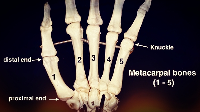
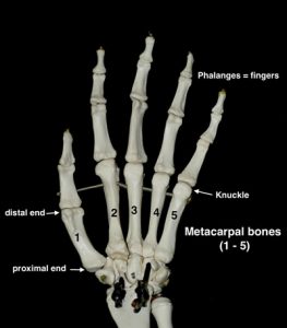
There are five bones in palm of your hand called metacarpals, one for each finger. The proximal end (situated nearer to the center of the body) of each metacarpal bone joins with the wrist. The distal ends (situated further away from the center of the body) forms the knuckles leading to the fingers. The five metacarpals fan out from the wrist towards the fingers. Each one is slightly different in shape and varies in degree of movement.
The metacarpals are easy to feel. Using your other hand explore them beginning with the thumb. Hold the thumb metacarpal as you bring your fingers together and spread them apart. Rotate the thumb in a small circle. Notice how moveable this bone is and how the proximal end curves around toward your palm. Your thumb is a rather unique design in that you can touch the pad of your thumb to those of your fingers. Only primates have this opposable thumb, which allows you to grasp objects and use tools.
Find the pinky finger metacarpal. Start at the knuckle (the distal end) and move toward the wrist feeling for the hardness of the bone. The proximal end is not as easy to define as the thumb metacarpal. The pinky metacarpal is the second most mobile of the five. While gently grasping this bone with your opposable thumb an index finger of your other hand wiggle the pinky metacarpal up and down and around in a small circle.
Continue to explore the three remaining metacarpals (index, middle and ring finger). They are less mobile than the thumb and pinky. The metacarpal associated with the index finger being the least moveable. Spread your fingers apart and notice how much each metacarpal moves laterally. Bring all your fingertips together while feeling how the metacarpals fold together to form cup. The thumb and pinky metacarpals move the most forming the sides of a cup while the other tree form the bottom.
The configuration of your metacarpals and wrist bones form three distinct hand arches. An arch is a curved structure spanning an opening. The proximal transverse (crosswise) arch forms along the line of carpometacarpal joints where the proximal end of the metacarpals meets the wrist bones. The distal transverse arch is along the line of the knuckles where the metacarpals meeting the phalanges (fingers). The longitudinal arch forms from the wrist to the fingers along the length of the palm. If you follow the 3rd metacarpal from the wrist toward the middle finger while your hand is cupped you can feel the shape of this arch. Explore the three arches of your palm as you flatten and cup your hand. Even when your palm is fully open you can still feel these arches.
 By comparison the horse’s anatomy in the corresponding area is quite simple. The front leg of the horse corresponds to your arm. Your wrist is his knee therefore the metacarpals are in the region between the knee and the fetlock joint. The cannon bone is considered as the 3rd metacarpal while the splint bones are the 2nd and 4th.
It is generally acknowledged that the ergot and chestnut are the remnants of the remaining two associated digits associated with the 1st and 5th metacarpals. These structures are like fingernails made of keratin, which would seem to be the end of the fingers but there aren’t any bones present. Therefore it makes one wonder what happened to all the bones associated with those two digits. I have heard a theory to explain what happened that I think is very plausible.
Jon Zahourek the inventor of Anatomy in Clay®and an avid comparative anatomist has proposed a theory to explain what happened to the five metacarpals. His theory is that the middle three metacarpals (index, middle and ring finger) joined together to form one bone – the cannon bone. The remaining two metacarpals correspond to the splint bones on either side. I think that this idea makes a lot of sense especially given the shape of the cannon bone condyles (rounded protuberance at the end of the bone forming an articulation with another bone) and the arch-like curve on the back side of the bone.
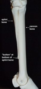
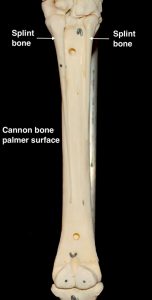 The splint bones are medial and lateral of the cannon bone toward the palmar (back) side. They are wider and thicker at the top and thin down toward the bottom of the cannon bone. They end approximately ¾ of the way down with a little button of bone that is easily palpable.
Next time you are with your horse run your hand down the cannon bone. Feel the rounded front surface. The back is filled with thick tendons so you can’t feel the curve of the bone on the palmer surface. Cup your hand around the back of the cannon bone with your thumb and index finger on the sides of the bone. Run your fingers down the sides of the following the splint bones. If you pay attention you will feel the buttons at the ends. Some horses have a calcification fusing the splint bone to the cannon.
Splints are generally caused by poor conformation or trauma to the leg. A splint is a calcification of the connective tissue between the cannon and the splint bone. Depending on the cause they horse may show signs of lameness as the splint is forming. Once they are “cold” they are considered a blemish are usually no cause for concern. If you feel a splint it is nothing to worry about if your horse is sound.
Wendy Murdoch resides in Washington, VA and is an international riding instructor/clinician. Wendy was a Pony Club Instructor for 6 years and continues to teach for Pony Clubs around the US. She travels worldwide teaching riders of all levels and disciplines how to improve the horse’s performance by improving their body position. Her book, 50 Five Minute Fixes to Improve Your Riding and Ride like a Natural 3 DVD set are available at www.murdochmethod.com.
[ux_custom_products cat=”magazine-subscriptions” products=”” columns=”4″ title=”Enjoy this article? Subscribe today!”]